The finger is conventionally divided into the region of the first and second phalanges, the region of the third phalanx, the region of the hoof sole and the pulp.
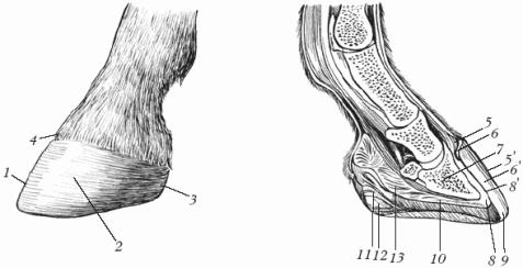
Area of the first and second phalanges. In this area of the finger, seven layers of tissue are distinguished (Fig. 1).
First layer-skin I. On the dorsal surface, the skin of the finger is connected to the deeper tissues (tendons and ligaments) rather loosely. From the volar surface, at the level of the border of the lower and middle third of the fetlock bone (above the crumb), the skin in the form of a jumper is firmly connected to the fascia of the finger.
Second layer- subcutaneous tissue. Functionally important blood vessels pass through this layer. In particular, 12-16 mm above the horny border, parallel to it, is the encircling (coronary) artery, which plays a significant role in the blood supply to the base of the skin of the corolla.
Third layer- thin superficial fascia. It reaches the fetlock joint, where it is lost in the loose subcutaneous layer.
Fourth layer- subfascial fiber. It loosely connects the thin superficial fascia with deeper tissues.
Fifth layer- deep fascia of the finger, is a continuation of the fascia of the forearm. The latter, having secured itself along the edges of the slate bones, passes to the area of the finger, where it covers the vessels and nerves, and on the dorsal surface it is very thin and merges here with the tendon of the common (long) extensor of the finger and ligaments. On the volar surface of the finger, the deep fascia is developed and reinforced by the lamellar ligament (which is sometimes called the fetlock fascia, or lamellar bandage). The latter, in the region of the first Phalanx, holds the superficial flexor tendon, anchored by two upper and two lower legs on the corresponding sides of the fetlock.
The deep fascia of the finger, covering the tendons of the common (long) extensor of the finger and the dorsal (accessory) branches of the interosseous middle muscle, connects with them, and also partially with the superficial flexor of the finger (I. Kolda).
Sixth layer- tendons, ligaments, vessels and nerves, which are located in this layer in the following order.
1. Tendons lie on the anterior (dorsal) and posterior (volar) surfaces of the finger. Here are the tendons of the common (long) extensor of the finger, the superficial and deep flexor of the finger.
Common (long) extensor tendon finger 2 runs along the dorsal surface of the finger, gradually widens and becomes thinner downwards, ending on the coronoid process of the coffin bone. At the level of the lower third of the second phalanx, the named tendon is quite tightly connected to the capsular ligament of the coffin joint.
Rice. 1. Horse’s finger from the lateral surface (copy from Kuznetsov’s dioptrogram):
1 - leather; 2 - tendon of the common extensor finger; 3 - fetlock bone; 4 - dorsal artery of the first phalanx; 5 - dorsal branch of the volar nerve; 6 - coronoid bone; 7 - dorsal artery of the second phalanx; B - anterior synovial eversion of the coffin joint; 9 - base of the corolla skin; 10 - coffin bone; 11 - base of the skin of the hoof wall; 12 - volar branch of the velar nerve; 13 - volar digital arteries and vein; 14 - supporting ligament; 15 - intermediate branch of the volar nerve; 16 - superficial digital flexor tendon; 17 - deep digital flexor tendon; 18 - posterior synovial eversion of the coffin joint; 19 - vein and artery arrows; 20 - navicular bone; 21 - soft cartilage; 22 - dorsal artery of the third phalanx and the nerve of the same name
Superficial digital flexor tendon 16 lies on the volar surface under the fascia. At the bottom of the metacarpus, this tendon wraps around the deep digital flexor tendon in a belt-like fashion. Then, going lower, in the area of the volar surface of the first phalanx, it divides into two legs and passes the tendon of the deep flexor of the finger between them. Each of these legs (lateral and medial) ends on the corresponding ligamentous tubercles of the coronoid and partly on the distal end of the fetlock. The named tendon is firmly fixed in the area of the sesamoid bones of the first phalanx by an annular ligament, and in the area of the volar surface of the fetlock bone by an x-shaped lamellar bandage (volar fetlock fascia), which is strengthened by two upper and two lower legs on the sides of this bone.
Flexor digitorum profundus tendon also lies on the volar surface of the finger. This tendon, passing between the terminal legs of the tendon of the superficial flexor of the finger above the navicular bone, greatly expands and covers the said bone in the form of a thin plate. Subsequently, the tendon of the deep flexor of the finger goes between the branches of the coffin bone and is attached in a fan-shaped manner to the tendon surface of the coffin bone and partially to the spinal cartilages.
The tendons of the superficial and deep flexor fingers have a common tendon sheath, which begins slightly below the middle of the metacarpus and ends at the level of the middle of the coronoid bone (N.V. Sadovsky).
2. Ligaments lie on the lateral (lateral and medial) and volar surfaces of the finger. This layer contains the lateral, intersesamoid, rectus, oblique and cruciate ligaments, as well as the interosseous muscle, which acts as a ligament.
Lateral, or collateral, ligaments (lateral and medial) begin on the lateral surfaces of the corresponding sesamoid bones. Each of them, divided into two legs, is fixed on the rough areas of the metacarpal (metatarsal) and fetlock bones.
Intersesamoid ligament connects the sesamoid bones to each other, forms a groove along which the tendon of the deep flexor of the finger slides.
The straight ligament originates from the base of the fetlock bones and is secured by deep fascicles at the base of the triangular roughness of the fetlock. The superficial bundles of the ligament terminate in a rough thickening of the coronoid bone.
The oblique ligaments are located along the edges of the previous ligament and also begin at the bases of the sesamoid bones and, converging with each other, end at the rough lines of the fetlock.
Cruciate ligaments located under the straight and oblique ligaments. Each of them originates from the base of the sesamoid bone, is directed obliquely downwards and, crossing with the ligament of the same name, is attached to the ligamentous tubercle of the fetlock on the opposite side.
Interosseous, or third (middle), muscle is built from tendon tissue. It begins on the volar thickened wall of the carpal joint capsule. In the distal third of the metacarpus it is divided into two branches, which end on the lateral surfaces of the sesamoid bones. Additional branches run from these branches to the common digital extensor tendon.
3. Vessels in this layer are represented by digital arteries and veins of the same name.
The digital arteries are located volar to the veins of the same name and descend along and parallel to the lateral (medial) edge of the deep digital flexor tendon. On their way, they give off the dorsal and volar arteries of the first phalanx 4, the artery of the pulp, the dorsal and volar arteries of the second phalanx 7.
The digital veins pass next to and dorsal to the arteries of the same name. At the level of the middle of the fetlock, the dorsal and volar veins of the second phalanx flow into them.
Lymphatic vessels are located in the subcutaneous tissue of the fingers near blood vessels and nerves (V. G. Martynov, I. V. Borodynya, P. F. Sorokova).
4. Nerves pass next to the vessels, forming neurovascular bundles. This layer contains the volar digital nerves (lateral and medial), which are divided above the fetlock joint into the dorsal and volar branches.
Dorsal branch of the volar nerve 5 has a very short trunk, which already at the level of the fetlock joint branches into two or three branches: one or two anterior and one intermediate. The anterior branches are located in front of the digital vein on the lateral surface of the first phalanx, and the intermediate branch of the volar nerve 15 passes between the volar digital artery and vein 13. These branches branch in the dorsal part of the skin of the finger, at the base of the skin of the hoof border and corolla, and in the anterior synovial eversion of the coronary and coffin joints (V.I. Troshin), and, in addition, partially take part in the innervation of the base of the skin of the hoof wall and the spinal cartilage (G. Nerper).
Volar branch of the volar nerve 12 is located along the posterior edge of the own artery of the finger. Descending down the lateral surface of the tendon of the superficial flexor of the finger and partially of the tendon sheath of the flexor of the finger, the volar branch at the level of the proximal edge of the spinal cartilage gives off a branch for it, passes to the inner surface of the said cartilage and along it reaches the plantar foramen coffin bone, where it plunges into the terminal (lunar) canal of the third phalanx, merging with the nerve of the same name on the opposite side (B. M, Olivekov).
According to V.I. Troshin, A.F. Ryzhikh, the final part of the volar nerve (in all cases) along the vascular notch of the lateral process (branch) of the coffin bone reaches the dorsal surface of the latter and branches at the base of the skin of the hoof wall.
In addition, the volar branches of the volar nerves take part in the innervation of the volar surface of the finger, the superficial and deep tendons of the flexor muscles of the finger and their tendon sheath, the posterior synovial eversion of the fetlock, coronary and hoof joints, the shuttle mucosa of the bursa and the volar part of the base of the skin of the hoof.
The dorsal and volar branches of the volar nerves have a number of connecting branches (V.I. Troshin).
On the pelvic limbs, the plantar nerves basically have the same anatomical and topographical position as the volar nerves. Each plantar nerve above the fetlock joint (in its most common form) is divided into two branches; in other, rare cases, the plantar nerve is not divided into digital nerves, and two or three dorsal digital branches extend from its trunk (A.F. Ryzhikh).
Seventh layer - supporting elements of the finger, which includes the fetlock, coronoid, coffin and navicular bones, as well as the joints formed by the articulation of these bones. There are 3 joints on the finger: fetlock, coronoid and ungulate.
The fetlock joint (the joint of the first phalanx) is formed by the articulation of the metacarpal (metatarsal), fetlock and two sesamoid bones. The joint capsule connecting these bones is fixed 2-3 cm above the articular surfaces of the metacarpal bone and along the articular surfaces of the first phalanx and sesamoid bones. This capsule forms two synovial eversion: the anterior one is small, quite closely connected with the tendon of the common (long) extensor finger, and the posterior one, much larger, located directly above the sesamoid bones. The posterior synovial eversion extends superiorly to the button-like thickening of the slate bones, and volarly to the anterior edge of the interosseous muscle. The fetlock joint also has two lateral ligaments (lateral and medial), closely connected to its capsule. They begin in the ligamentous fossae of the third metacarpal bone and end on the ligamentous tuberosities of the first phalanx.
The coronoid joint (the joint of the second phalanx) is formed by the articulation of the fetlock and coronoid bones. The joint capsule has two synovial eversion. The anterior eversion is located under the tendon of the common (long) extensor of the finger and is closely connected to it. Its lower border is located 3-4 cm above the horny capsule. The posterior synovial eversion on the volar surface is covered by the deep digital flexor tendon. The collateral ligaments are partially intertwined with the fibers of adjacent fibrous tissue. In addition to these ligaments, the coronoid joint additionally has two pairs of short, strong volar ligaments (two middle and two lateral volar ligaments), which begin on the volar surface of the fetlock and end at the proximal end of the volar surface of the coronoid.
The coffin joint (the joint of the third phalanx) is formed by the articulation of the coronoid, coffin and navicular bones. The articular capsule is attached to all bones and has two synovial eversions. Anterior synovial eversion 8 is located under the tendon of the common (long) extensor finger. In the lower two-thirds of its length, the anterior eversion is closely connected to the indicated tendon. With its posterior wall it is adjacent to the dorsal surface of the coronoid bone, extending to two-thirds of its height. The upper part of the anterior inversion is not a continuous protrusion, but consists of a number of additional inversions (from 2 to 5), located at a short distance from each other.
Posterior synovial eversion of the coffin joint 18 in the filled state also reaches the level of the border of the middle and upper third of the volar surface of the coronoid bone. It, in turn, has a number of small additional protrusions. These protrusions are directed upward to the volar roughness, without reaching the proximal end of the bone of the second phalanx.
Collateral ligaments of the coffin joint they begin in the ligamentous fossae of the second phalanx and, expanding fan-shaped, are attached to the coffin bone, volar to its extensor process.
Region of the third phalanx. This area is divided into 3 areas: the hoof border, the hoof crown and the hoof wall. There are five layers here.
The first layer is the horny border. It is located at the border of the skin and the horn capsule in the form of a narrow (3-5 mm wide) and thin (0.5-0.8 mm thick) strip. The horn border consists of a light gray soft horn, which, falling down onto the hoof wall, forms its superficial (glaze) layer.
Below the horny border is the horny wall of the hoof. The thickness of the horny wall at the toe reaches 8-10 mm, towards the heel parts the wall becomes thinner (up to 6 mm), and on the hind hooves it is slightly thicker. The horny wall of the hoof, in turn, consists of three layers: the superficial - glazed, middle - tubular horn and deep - leafy horn. The latter is directly adjacent to the producing layer of the epidermis.
The proximal edge of the horny wall, together with the horny border, has a groove on its inner surface (1-1.5 cm wide and up to 1 cm deep). The hoof corolla is located in this groove.

Rice. 2. Base of leather: borders; 2 - corolla; 3 - hoof stakes

Rice. 3. Pulmonary (hoof) cartilage 1 - coronoid bone; 2, 4 - cartilage ligaments; 3 - coffin bone: 5 - spinal cartilage
The second layer is the base of the skin (Fig. 2). From top to bottom, starting from the skin, this layer is anatomically divided into: the base of the skin border / (3-5 mm wide); base of corolla skin 2 (width 1-1.5 cm); base of the skin of the hoof wall 3.
The base of the skin consists of an inner vascular layer and an outer layer, which in the area of the hoof border and hoof corolla has a papillary structure, and in the area of the hoof wall has a leaflet structure.
At the base of the skin there is a dense network of arterial, venous and lymphatic vessels.
The most developed nerve bundles are located in the periosteal layer, from where they are directed to the vascular layer, forming a dense network. Next, they penetrate into the leaflet layer, where the nerve bundles are located along the entire length of the base of the leaflet (V.S. Dudenko).
The third layer is the subcutaneous layer. It consists of fairly loose connective tissue. Its thickness in the area of the hoof border is up to 1 mm. In the area of the hoof rim this layer looks like a 1.5 cm thick roller; in the area of the hoof wall it is completely absent. Here the base of the skin of the hoof wall is directly connected to the coffin bone, and volarly to the spinal cartilage.
Fourth layer - distal part of the coronoid and the coffin bone in the area of the toe wall of the hoof and the spinal cartilages (lateral and medial) in the area of the lateral walls of the hoof.
The soft cartilage (Fig. 3) has the appearance of a somewhat elongated elastic diamond-shaped plate with rounded corners. Its thickness in the upper toe part is 0.3-0.7 cm, and towards the heel parts it thickens like a club.
The lower border of the anterior third of the cartilage descends under the horny border to 1.3-1.9 cm, and the posterior third - to 1.5-2.5 cm. The upper border of the cartilage protrudes above the horny capsule up to 2.5 cm and gradually decreases towards toe and heel parts (P. N. Skvortsov).
The anterior elongated part of the said cartilage with its inner surface covers almost the entire lateral ligament of the hoof joint and is quite firmly connected to it, its middle part is adjacent to the posterior synovial eversion of the said joint, and the volar part is adjacent to the crumb (F. Richard).
The soft cartilages are connected by ligaments to all the bones of the finger. They have two (internal and external) venous networks connected to each other by anastomoses. When performing the operation, it must be borne in mind that directly behind the volar surface of the coronoid bone on the inner side of the spinal cartilage lie: the dorsal artery of the second phalanx, the main highways of the volar (ilantar) artery and vein, the volar (ilantar) branch of the volar (plantar) nerve, which are adjacent to the lateral surface of the posterior synovial eversion of the coffin joint. Therefore, from a practical standpoint, it is important to remember that any deep cuts in this area will be accompanied by severe bleeding, as well as disruption of the integrity of functionally important nerves and the capsule of the hoof joint.
Fifth layer - extensor process of the coffin bone in the toe area of the hoof, the anterior third of the spinal cartilage, the lateral ligaments of the coffin joint and the coronoid bone in the area of the lateral walls of the hoof. In the posterior half of the spinal cartilage, in the fifth layer there is a crumb; the latter fills the space limited by the spinal cartilages (lateral and medial) and the posterior (volar, plantar) surface of the deep digital flexor tendon. On the volar surface, the crumb has the following layers: the horn of the crumb is 1-2 mm thick, producing a layer of epidermis and the base of the skin 2-4 mm thick, a subcutaneous layer 2-2.5 cm thick (N.V. Sadovsky).
Hoof sole area(Fig. 4). The hoof sole is the lower (supporting) part of the horn capsule. The place where it connects with the horny wall, expressed in the form of a light semiring, is called the white line 3.
In single-hoofed animals (horses, donkeys, mules), the heel walls in the back of the hoof curl forward, forming a bar angle (column) 1 and then move to the sole, where they are called bar walls 8. Between the bar walls there is a horny arrow 6. On it in the middle there is a mid-arrow groove 7. Between the outer edges of the frog and the bars there are lateral (lateral and medial) grooves of the frog 2.
In the sole area, three layers are distinguished (Fig. 4).
The first layer is the sole horn 10, 6-8 mm thick, consisting of horny tubes.
The second layer is the producing layer of the epidermis and the basis of the skin of the sole. The latter has a thickness of 4-6 mm and consists of the papillary and vascular layers. Near its peripheral edge, adjacent to the hoof wall, there are the girdle artery and vein of the sole 3.

Rice. 4. Horse hoof (view from the sole): 1- bar angle (column); 2 - lateral groove of the arrow: 3 - white line; 4 - plantar edge of the horny wall: 5 - horny sole; 6 - horny arrow; 7 - mid-arrow groove: 8 - bar wall

Rice. 5. Horse hoof from the plantar surface (copy from Kuznetsov’s diopttrogram):
1- artery and vein arrows; 2 - accessory artery of the sole; 3 - girdle artery and vein of the sole; 4 - arterial and venous network of the sole; 5 - base of the skin of the hoof wall; 6~ projection of the navicular bone; 7 - projection of the peripheral edge of the coffin bone; 8 - gate walls 9 - white line; 10 - sole horn
The third layer is the coffin bone. The projection of the peripheral edge of this bone 7 almost coincides with the projection of the same edge of the base of the skin of the hoof wall 5 and lies 4-6 mm to the center from it.
Area of the hoof arrow. The hoof arrow has the shape of a wedge, the top of which is directed towards the toe (see Fig. 4). It is limited by the calcaneal branches of the sole and the bars. In the area of the hoof arrow, eight layers are distinguished.
The first (outer) layer is soft elastic horn thickness 7.5-15 mm.
Second layer - the producing layer of the epidermis and the base of the skin of the arrow. The latter has papillary and vascular layers, the thickness of which is 2-4 mm.
Third layer - subcutaneous (subcutaneous arrow), consists of fibrous tissue and elastic fibers. The subcutaneous arrow at its base is up to 1.5 cm thick; closer to the top of the horny arrow, the thickness of its subcutaneous layer gradually decreases, and at the very top of the arrow, this layer disappears completely. The lateral and medial arteries, veins and nerves of the arrow pass through this layer (see Fig. 5). The named vessels and nerves lie at the level of the middle of the legs of the arrow (G. S. Kuznetsov).
Fourth layer - cruciate ligaments of the spinal cartilages. Each of these ligaments begins on the inner surface of the spinal cartilage and ends on the opposite lateral process (branch) of the coffin bone and partially on its semilunar crest. The cruciate ligaments are firmly connected to the subcutaneous layer of the pulp.
Fifth layer-- plantar fascia. It begins with two legs in the ligamentous fossae of the distal end of the fetlock. These legs soon after their onset fuse in an arcuate manner and form the plantar fascia; the latter covers the outside of the tendon of the deep flexor of the finger and ends on the semilunar crest of the coffin bone. The terminal part of the plantar fascia, from its insertion to the lunate crest of the coffin bone and to the level of half of the navicular bone, is quite firmly connected to the tendon of the deep digital flexor. Above and volarium of the navicular bone, the plantar fascia connects with the named tendon more loosely.
Sixth layer - terminal portion of the deep digital flexor tendon. In this layer it passes between the spinal cartilages. In the area of the navicular bone, the tendon expands, thins, becomes flat and completely covers the said bone. Here the deep digital flexor tendon is most poorly vascularized.

Rice. 6. Horse hoof in plantar projection (copy from dioptrogram according to Kuznetsov):
1 - shuttle mucous bursa; 2 - plantar (digital) artery and vein; 3 - coffin bone; 4 - base of the skin of the hoof wall; 5- projection of the navicular bone
Seventh layer - shuttle mucous bursa(Fig. 6, 1). When filled, it resembles the shape of an irregular semicircle with wavy edges. In some cases, at the level of the lateral edges of the navicular bone, this bursa has additional (lateral and medial) protrusions reaching the size of a kidney bean. The named mucous bursa is somewhat (2-3 mm) wide beyond the ends (lateral and medial) of the navicular bone 5 and extends from it upward and backward by 10-12 mm. The size of the shuttle mucous bursa in adult horses reaches a length (perpendicular to the axis of the bone of the same name) up to 3.6 cm, and a width of up to 5.6 cm. The shuttle mucous bursa from the frog side is completely covered by the tendon of the deep flexor of the digitalis, with the exception of additional protrusions, which are 5-7 mm volar to the sesamoid bone of the third phalanx and extend beyond the edges of the tendon.
Between the lateral edge of the shuttle mucous bursa and the lunate line of the coffin bone 3, the digital (plantar, according to B. M. Olivekov) artery and vein 2 pass in the loose tissue. This anatomical position should be kept in mind when resection of the deep digital flexor tendon in order to avoid damage to these vessels, which are essential in the blood supply to the coffin bone. As an exception, the shuttle bursa can communicate with the digital tendon sheath (B. M. Olivekov), as well as with the cavity of the hoof joint (N. Z. Nemirovsky).
Eighth layer - coffin and navicular bones, connected to each other in this area by the hoof-shuttle ligament. The latter is closely connected with the articular capsule of the coffin joint. Separate bundles extend from the hoof-shuttle ligament, forming a very thin unpaired ligament. From the posterosuperior edge of the navicular bone originates the suspensory ligament of the navicular bone, which covers the posterior synovial eversion of the coffin joint from the volar (plantar) surface. This ligament ends on the lateral surfaces of the fetlock bone.
The posterosuperior edge of the navicular bone usually lies on the line connecting the ends of the rami of the sole. Therefore, it is rational to make a transverse incision of the arrow during partial resection of the tendon of the deep flexor of the finger according to the indicated projection so that at the subsequent moment of the operation, stepping back 5.5 mm dorsally (towards the toe), it is possible to cut the said tendon within the boundaries of the navicular bone. With this incision, accidental damage to the navicular ligament, the suspensory ligament of the navicular bone and the capsular ligament of the coffin joint closely adjacent to them, as well as the arteries and veins involved in the blood supply to the navicular bone, are excluded.
Closely associated with the hoof-natchicular ligament, the inferior synovial eversion of the coffin joint is located 16-20 mm (the width of a finger) dorsal to the line connecting the ends of the branches of the sole, therefore, when opening this eversion there is no need to resect the top of the hoof frog. It is more advantageous to open the joint itself in the middle of the hoof-natchetal ligament, which avoids damage to the volar transverse arches of the artery and vein of the third phalanx (G. S. Kuznetsov).
Burdenkzh A.F. and Kuznetsov G.S. Veterinary orthopedics.
Pulvini are dense, pillow-shaped thickenings of skin on the back surface of the paw. Horses have carpal and tarsal pads (chestnuts), metacarpal and metatarsal pads (spurs), and digital pads.
Chestnut located on the medial surface of the distal end of the forearm and tarsus, spur- on the back surface of the 1st phalanx of the finger and covered with a brush. Chestnuts and spurs are hairless areas of the skin. Consist of epidermis, dermis and subcutaneous tissue. The epidermis is thick, highly keratinized, and forms soft keratin. Cattle and pigs only have finger crumbs.
The finger pulps are located on the distal phalanx of each finger. Consist of epidermis, dermis and subcutaneous tissue. The epidermis is highly keratinized, consists of five layers, its thick stratum corneum is formed by a tubular horn. In the dermis there are papillary and reticular layers, many elastic fibers, blood vessels and sweat glands. A large number of nerve endings makes the digital pulp an organ of touch. In the subcutaneous layer, a cushion of crumb is formed from fat accumulations, which acts as a shock absorber. It is especially developed in its posterior part.
In horses, the toe pads have the shape of a triangle forked at the base, wedging its apex into the sole of the hoof. Its rear, more elastic part forms a cushion of the crumb, and its sharp, elastic front part forms a frog with a massive stratum corneum. IN arrow distinguish the apex, legs, interpeduncular groove, and on the inside - the ridge of the arrow. The horse has paired pulpal cartilages fused with the cushion of the pulp. Cartilage performs a spring function. In cattle and pigs, the finger crumbs do not have arrows.
Fig.1. Horse hoof
1 – horny pillow; 2 – medial leg of the arrow; 3 – tip of the arrow; 4 – interpeduncular groove; 5 – lateral groove of the arrow; 6 – plantar edge of the hoof wall; 7 – horny pad of the hoof; 8 – leg of the sole; 9 – collar part of the hoof; 10 – inversion angle of the hoof; 11 – white line of the hoof; 12 – base of the skin of the hoof border; 13 – base of the skin of the hoof corolla; 14 – base of the skin of the hoof wall; 15 – hoof contour.
Hoof– ungula – is divided into the hoof border, hoof corolla, hoof wall and hoof sole. Hoof border– looks like a narrow strip, about 0.5 cm wide. Consists of the epidermis, dermis and subcutaneous layer. The epidermis of the border has all five layers (basal, spinous, granular, lucid and horny). The stratum corneum of the epidermis forms the glaze of the hoof wall. The dermis consists of the papillary and reticular layers and contains many blood vessels. The subcutaneous tissue is slightly developed and passes into the periosteum of the finger.
Hoof corolla– about 1.5 cm wide, located below the hoof border. Consists of the epidermis, dermis and a faint subcutaneous layer. In the epidermis, the germinal zone immediately transitions into the thick stratum corneum, which produces the thick tubular horn of the hoof wall. This horn is very strong, consists of horn tubes in which the cells are tightly adjacent to each other. The tubes are soldered together by an intermediate horn. The tubular horn is pigmented. The dermis is highly developed and hangs in the form of a coronary ridge over the wall of the hoof. A coronary groove is formed from the inside. There are many vessels in the dermis. The subcutaneous tissue passes into the periosteum of the finger.
Hoof wall- the most massive part of the hoof. It distinguishes between an unpaired toe, lateral medial, lateral lateral parts, inversion angles, and plantar edge. It consists of the epidermis and dermis, there is no subcutaneous layer. The epidermis produces a white leaf horn that forms a white line on the sole of the hoof. The white line indicates the boundary beyond which one should not go when shoeing a horse, so as not to damage the living tissue of the finger.
Sole– consists of epidermis and dermis. The epidermis forms a tubular horn. The dermis passes into the periosteum of the third phalanx of the finger. On the sole of the hoof, a body and plantar branches are distinguished, between which a horny digital crumb with an arrow is wedged.
Kopytse– ungulicula – in cattle and pigs it consists of the same parts as in the horse (edge, corolla, wall and sole). However, the body and plantar branches are not distinguished on the sole, and the arrow is missing on the digital crumb.
Thus, horn capsule hooves and hooves have a complex structure. Its wall consists of three layers. The most superficial layer is glaze. It is thin and breaks down quickly. Middle layer - tubular horn. It is the thickest and most durable, and contains pigment that gives it a dark color. Inner layer - leaf horn, does not contain pigment. All layers of the cole horn are clearly visible when trimming hooves or hooves. The horn capsule normally grows at the same rate as it is erased. Metabolic disorders lead to excessive keratinization. Dietary disorders can cause the shoe to slow down and reduce its elasticity. At the same time, grooves appear on the hooves and hooves, and their surface becomes dull and rough.
Horn– cornu – heavily keratinized skin cover of the horny process of the frontal bone of the skull. The horn is divided into root, body and apex. The horn is formed by the epidermis and dermis. The dermis is formed by the papillary and reticular layers. The reticular layer passes into the periosteum of the corneal process. The epidermis produces a strong tubular horn. The horn grows intensively at a young age. In the cold season, with insufficient feeding and during pregnancy, horn growth slows down, which leads to the appearance of rings, noticeable in cattle near the root, and in sheep and goats throughout the horn. The approximate age of a cow can be determined from the horn rings by adding the number 2 to the number of rings.
The structure of the mammary glandsBreast– glandula lactiferae – is a characteristic feature of the class of mammals. It reaches its maximum development in females before the birth of the baby during lactation. In different animal species, milk jellies have different shapes, sizes, location and composition of secreted secretions. They can be located in the form of one pair of hills in the chest area (in elephants, primates and cetaceans), in the groin area between the thighs (in horses and cattle). In omnivores, rodents and carnivores, the mammary glands in the form of isolated separate hills are located on both sides along the linea alba of the abdomen. The mammary gland of farm animals is called the udder.
Udder– uber – large, complex, alveolar-tubular gland. In cattle and horses, the udder is simple, its lobes merge into a single organ located in the groin area between the thighs. In pigs, the udder is multiple, located along the linea alba in the form of 4-8 pairs of milk mounds lying on the ventral surface of the chest and abdomen. Cattle and horses have 4 udder lobes, sheep and goats have 2 lobes. In cattle, each lobe has its own nipple. In horses and pigs, one teat is connected to two lobes.
The surface of the udder adjacent to the abdomen is called base of the udder, the entire mass of the gland is udder body, the ventral part of the organ between the nipples – udder bottom. Caudal folded surface of the udder - milky mirror. Depending on the shape, the udder is cup-shaped, bath-shaped, round, flat and goat-shaped. The ideal shapes for machine milking are bath-shaped and bowl-shaped. According to the position, the udder can be femoral (displaced in the caudal direction) and abdominal (displaced in the cranial direction). The surface of the udder is covered with skin with sweat and sebaceous glands and sparse, delicate hair. Under the skin is superficial fascia, and under it deep fascia. It is a continuation of the yellow abdominal fascia with a large number of elastic fibers. The deep fascia forms suspensory ligament, which divides the udder into left and right halves. Each half consists of two lobes: anterior and posterior. Each lobe has its own system of ducts and its own nipple. The nipple is distinguished base, body And top. There are three types of nipples: cylindrical, conical and teardrop-shaped (pear-shaped). Cylindrical teats are most suitable for machine milking. The skin of the nipples is hairless, there are no sebaceous and sweat glands, but there are many sensitive nerve endings.
Under the deep fascia there is a connective tissue capsule, from which connective tissue partitions - trabeculae - extend deep into the organ. The capsule and trabeculae form the skeleton of the udder or stroma. Elements of the stroma do not perform specific functions, that is, they do not synthesize milk. Blood and lymphatic vessels, milk ducts, nerves pass through the stroma, and accumulations of fat cells are deposited. The glandular part of the udder - parenchyma. It is formed by glandular epithelium, from which the end sections of the mammary gland and the system of excretory ducts are built. Trabeculae penetrate deep into the organ and divide the parenchyma into lobules. Lobules are formed alveolar tubes. The alveolar tube wall is formed by two layers. The inner layer consists of a single-layer cubic or columnar glandular epithelium. Each cell secretes all the components of milk. Secretion droplets are released according to the merocrine or apocrine type of secretion. The outer layer of the alveolar tubes is formed by branched myoepithelial or “basket” cells. They are able to contract and compress the alveolar tubes, which helps to release milk. The udder is penetrated by vessels, which are branches of the external pudendal artery. Capillaries densely entwine each alveolar tube.
From the alveolar tubes milk flows into intralobular excretory ducts. Their diameter is smaller than the alveolar tubes, and the wall is lined with single-layer cubic epithelium. Intralobular excretory ducts unite into interlobular excretory ducts - milk ducts, and these, in turn, merge into wide milk passages, clearly visible to the naked eye. The wall of the mammary ducts is lined with double-layer epithelium. Milk passages open in milk tank. Each lobe of the udder has its own milk tank. It is an irregularly shaped cavity with a volume of up to 100-120 ml. The milk tank opens in teat cistern, which turns into a short nipple canal, opening outwards. The mucous membrane of the nipple canal is formed by stratified squamous epithelium. The wall of the nipple contains smooth muscle tissue. In the area of the nipple canal, the annular muscle layer forms the sphincter of the nipple.
A horse's udder has two milk mounds and two teats. Each nipple is connected to two milk lobes and contains two teat cisterns and two nipple canals. Cattle have four milk mounds and four teats. Each nipple has one teat cistern and one teat canal. Small cattle have two milk mounds, two teats containing one tank and one canal. Pigs have 1-3 cisterns and the same number of channels in each teat.
The influence of the physiological state of the animal on the structure of the udder
The mammary gland reaches its highest functional activity during lactation. At this time, the parenchyma makes up 70-80% of the mass of the gland. The alveolar tubes are closely adjacent to each other, have a wide lumen, high epithelium and delicate connective tissue layers of loose fibrous tissue. By the end of lactation, the size of the alveolar tubes decreases, and the intralobular and interlobular connective tissue layers of fibrous tissue thicken. Clusters of fat cells appear in them. During the dry period, the end sections of the gland collapse, the epithelium becomes low, the connective tissue layers are wide with significant fatty deposits. In old and unproductive animals, the stroma is better developed than the parenchyma. The connective tissue of the stroma is denser than in highly productive animals. In well-fed animals, the stroma increases due to the deposition of fat in the interlobular connective tissue layers and under the skin of the udder.
Section three. Orthopedics. Chapter XXVI. ANATOMY AND FUNCTION OF THE HOOF ANATOMY OF THE HOOF
The hoof (Ungula) is a skin transformed at the end of the toe into a hard skin tip. According to this definition, the composition of the hoof should include only those tissues that are characteristic of the skin, i.e., the subcutaneous layer, the base of the skin and the epidermis. Bones, ligaments, tendon endings of the muscles enclosed in the hoof belong to the organs of movement. However, to substantiate the rules of shoeing, correct orientation in hoof diseases and their treatment, knowledge of the structure of not only the hoof, but also the finger as a whole is required.
The structure of the cattle claw. Cattle have two separate functioning fingers - the third and fourth, and two rudimentary ones - the second and fifth (Fig. 138).
The skeleton of each main finger consists of three phalanges and three sesamoid bones.
The bone of the third phalanx, or claw bone, looks like a slightly curved triangular pyramid, the apex of which is directed forward (Fig. 139). It has three surfaces: articular, two steppe (external and internal) and plantar.
On the dorsal side of the articular surface there is an extensor process for attaching the tendon of the common (long) extensor digitorum, and on the volar side there is a flexor process for attaching the tendon of the deep flexor digitorum.
Rice. 138. Sagittal section of the toe of the thoracic limb of cattle:
The hoof is divided into five anatomical parts: 1) the hoof border; 2) hoof corolla; 3) hoof wall; 4) hoof sole; 5) finger crumb.
The claw border looks like a strip 4-7 mm wide. It surrounds the finger in a ring shape at the upper contour of the horny capsule, and at its volar surface it passes into the crumb; the hoof border consists of the epidermis, the base of the skin and the subcutaneous layer. The base of the skin of the hoof border is slightly convex and covered with papillae.
The functional significance of the hoof border is as follows.
1. The producing layer of the epidermis of the hoof border produces a soft horn - the horny border, which, falling onto the hoof wall, forms a thin shiny layer - the glaze of the hoof wall.
2. The hoof border connects the horny shoe with the hairy skin, relieves the pressure of the coronal edge of the horny capsule on the adjacent area of the skin, serves as a kind of hoop, covering the horny capsule at the top and ensuring the preservation of its contour.
The hoof corolla is about 2-2.5 cm wide, located in a semi-ring below the hoof border. It, like the hoof border, consists of the epidermis, the base of the skin and the subcutaneous layer. On the base of the skin of the hoof corolla there are numerous papillae up to 1.8 cm long.
The functional significance of the hoof corolla is as follows.
1. The producing layer of the epidermis of the hoof corolla forms a tubular horn, which, going down, forms the most powerful middle, or coronal, layer of the horny wall.
2. The highly developed subcutaneous layer of the hoof corolla somewhat softens shocks and shocks when the hoof rests on the ground and, to a certain extent, protects the tissue enclosed in the horny shoe from damage.
3. The hoof corolla serves as an organ of touch, thanks to which animals distinguish the nature of the soil when moving.

Rice. 139. Cattle hooves:
The hoof wall is represented by the epidermis with the stratum corneum and the base of the skin. There is an outer horny wall - convex and inner - slightly concave; both walls on the dorsal surface converge into a blunt edge. The wall thickness decreases from front to back; it reaches 5 mm in the middle of the inner wall and 7 mm in the outer wall. The base of the hoof wall is covered with a large number of leaves, which are relatively low, short and located in the lower half of the hoof wall (Fig. 140).
The functional significance of the hoof wall is as follows.
1. The hoof wall protects the base of the skin and underlying tissues of the claw from mechanical, physical and chemical damage.
2. The presence of a leafy horn at the base of the skin of the hoof wall provides a strong (“lock”) movable connection with the underlying tissues, in addition, it contributes to the uniform distribution of body weight throughout the entire claw and the softening of shocks and shocks that occur when leaning limbs on the ground.
3. The producing layer of the epidermis of the hoof wall produces horny leaves.
The hoof sole is very narrow; it passes posteriorly into the digital pulp. The hoof sole consists of the epidermis and the base of the skin. The base of the skin of the hoof sole is covered with leaves, which are a continuation of the leaves of the hoof wall.
The functional significance of the hoof sole is as follows.
1. The horny sole protects the underlying tissues from mechanical damage and participates in the formation of the horny shoe.
2. The producing layer of the epidermis of the sole of the claw produces a tubular horn, forming the horny sole.
The digital soft tissue is highly developed; its thick stratum corneum is made of soft tubular horn, and the subcutaneous layer, 1-1.5 cm thick, is made of strong collagen and elastic fibers. The base of the skin of the crumb is covered with small papillae.
The functional significance of the digital crumb is as follows.
1. The crumb serves as an elastic spring, softening shocks and shocks when a limb rests on the ground; In addition, it increases the area of support on the ground and prevents the hoof from slipping.
2. The finger crumb performs tactile functions in a certain part.
The blood supply to the fingers comes from numerous branches. On the thoracic limb, the fingers are supplied with blood from 4 sources: 1) the common dorsal digital artery, from which two special digital arteries are separated - the lateral fourth and medial third; 2) third common volar digital artery; 3) second common volar digital artery; 4) the fourth common volar digital artery. On the pelvic limb, the blood supply comes from 3 sources: 1) the common dorsal digital artery, which is divided into two special dorsal digital arteries of the third and fourth fingers; 2) the medial plantar artery, which above the fetlock joint is divided into two branches - the second and third plantar digital arteries; its first branch subsequently passes into the special plantar artery of the third finger;. 3) the lateral plantar artery, which passes into the special plantar artery of the fourth finger.

Rice. 140. Base of the skin of the hooves: /Rice. 141. Sheep interclaw gland (incision made between the 3rd and 4th toes): /Rice. 142. Pig fingers from the volar side:
The veins of the finger have the same name as the arteries.
The innervation of the finger on the thoracic limb is carried out through the branches of the median, radial and ulnar nerves, and on the pelvic limb - through the branches of the spinal and peroneal nerves.
Features of the anatomical structure of sheep's hooves. The hooves of sheep have the same anatomical structure as those of cattle, but with some features, the essence of which boils down to the following.
1. At the level of the coronary joints between the third and fourth fingers there are interdigital sacs, or interclaw glands (Fig. 141). They are skin depressions in the wall of which the sebaceous and glomerular glands lie.
The outlet of the interdigital sac with a diameter of 2-3 mm lies on the dorsal surface of the finger at the level of the coronoid joint.
2. The hoof horn is thin. The thickness of the outer horny wall is 2.5-3 mm, the inner one is 1.5-1.8 mm, the horny sole is 2.5-3.5 mm and the stratum corneum is 5 mm.
3. The sole of the hoof occupies only the front part of the plantar surface of the claw, the larger part, the back part of which is the digital crumb.
The blood supply to the sheep's fingers comes primarily from the common volar (plantar) digital artery and, to a lesser extent, from the dorsal middle metacarpal (plantar) artery.
The innervation of the finger is carried out by multiple (L-9) branches extending from the ulnar, radial and median nerves.
Features of the anatomical structure of the hooves of pigs. Pig hooves are divided into the same parts as cattle hooves, but have the following features.
1 The horny shoe is relatively thin, its thickness at the coronal edge is 0.5-1 mm, in the wall area - 3-4 mm, at the sole - 2-3 mm.
2. The crumb is highly developed, occupies more than half of the sole and is clearly demarcated from the latter (Fig. 142).
3 The base of the coronary skin occupies the entire upper half of the hoof wall. The base of the skin wall has 130-180 leaflets.
The structure of a horse's hoof. The skeleton of a horse's toe consists of the coronoid fetlock, the coffin bone and three sesamoid bones. The horny shoe contains only two bones - the ungulate, or third phalanx, and the shuttle (Fig. 143).

Rice. 143. Sagittal section of a horse’s hoof:

Rice. 144. Base of hoof skin: /Rice. 145. Hoof sole, pulp and frog:
The coffin bone, or third phalanx, has a spongy structure and is shaped like the hoof. It distinguishes between articular, wall and plantar surfaces, as well as two branches with which the spinal cartilages are firmly fused. The tendon of the common (thoracic limbs) and long (pelvic limbs) extensor finger is attached to the extensor process of the coffin bone, and the tendon of the deep flexor finger is attached to the plantar surface.
The shuttle bone has the shape of a weaver's shuttle, is located between the branches of the coffin bone, and serves as a block for the sliding of the tendons of the deep flexor of the finger. This is facilitated by the presence of a mucous bursa on it - the shuttle bursa.
For ease of study, the horse’s hoof is also divided into the following anatomical parts: 1) hoof border; 2) hoof corolla; 3) hoof wall; 4) hoof sole; 5) finger crumb.
The hoof border is located between the hairy skin and the upper edge of the horny shoe in the form of a hairless strip 5-6 mm wide. It covers the front and side walls of the hoof and merges with the crumb at the back.
The hoof border consists of the subcutaneous layer, the base of the skin (Fig. 144, /) and the epidermis with the producing and horny layers.
The hoof crown is located downward from the hoof border. On a hoof with a removed horny shoe, it is impossible to determine the lower border of the hoof corolla, since it is covered from the outside by the coronal edge of the horny wall.
The hoof corolla is represented by all three layers of skin. Its subcutaneous layer is highly developed and has a coarse fibrous structure. The base of the skin of the corolla (Fig. 144, 2) is penetrated by a significant number of blood vessels and nerves. Its surface is covered with thick and rather long papillae, which are clearly visible to the naked eye. The subcutaneous layer and the base of the skin of the corolla form an elastic roll of the hoof corolla 1 - 1.5 cm wide, which is clearly visible after removing the horny shoe.
The hoof wall covers the wall surface and branches of the coffin bone. It is represented by two layers: the base of the skin and the epidermis with the stratum corneum. There is no subcutaneous layer in the wall area.
The base of the skin wall, in turn, consists of three layers: periosteal, vascular and lamellar. The deepest periosteum of the base of the skin is firmly fused with the periosteum of the coffin bone. The outer layer of the base of the skin of the wall has a leaf-like structure (Fig. 144, 3). There are 500-600 leaves on the hoof wall.
The productive layer of the epidermis produces horny leaves, which in their shape and number correspond to the leaves of the base of the skin wall. The horny leaves form the deepest layer of the horny wall - the leaflet. Thus, the horny wall of the hoof consists of three horny layers: 1) superficial - glaze; 2) middle - coronal; 3) deep - leafy.
On the horny wall there are distinguished: toe, lateral, heel and turn parts, as well as coronal and plantar edges. The places where the horny wall bends onto the plantar surface are called the turn, or heel, angles.
The hoof sole (Fig. 145) occupies the lower surface of the hoof and consists of two layers: the base of the skin and the epidermis with the stratum corneum.
The base of the skin of the sole fuses with the periosteum of the plantar surface of the coffin bone. Its outer surface has a papillary structure. The producing layer of the epidermis (not visible to the eye, covers the papillae of the base of the skin of the sole) produces a tubular horn - the horny sole. The latter is a slightly concave horny plate with a cutout for the arrow.
On the horny sole there is a body (front part) and two branches. The ends of the branches form plantar angles.
The junction of the plantar edge of the horny wall with the sole is called the white line, which in appearance is a light yellowish strip approximately 4 mm wide. The white line is formed by the ends of the horny leaflets and the tubular horn that fills the spaces between the leaflets. The thickness of the horny wall can be determined by the white line.
The digital crumb lies between the bar walls and the spinal cartilages. It has the shape of a wedge, bifurcated by a longitudinal groove. On the finger crumb, a soft pad and a crumb arrow are distinguished.
The soft pad is the thickened posterior part of the digital crumb; the arrow of the crumb is its pointed front part.
The digital crumb consists of the subcutaneous layer, the base of the skin of the crumb and the epidermis with the producing stratum corneum.
The subcutaneous layer of the crumb is highly developed, containing collagen and elastic fibers and layers of adipose tissue. The base of the skin of the crumb has a papillary structure. The epidermis of the crumb produces a thick but soft stratum corneum, which in the region of the frog is called the horny frog.
On the horny frog there are distinguished: legs (femurs) of the frog, mid-arrow groove, lateral frog grooves, body and apex of the frog.
The functional significance of the anatomical parts of the horse's hoof discussed above is similar to the functional significance of the corresponding parts of the cattle's hoof (see structure of the cattle's hoof).
The blood supply to the hoof tissues is carried out by the branches of the volar (plantar) digital arteries, which on the thoracic limb are a continuation of the superficial volar metacarpal artery, and on the pelvic limb - the metatarsal dorsal lateral artery.
The digital arteries are located along the lateral and medial edges of the deep digital flexor tendon, go down to the plantar foramen of the coffin bone, enter the semilunar canal on each side, where they form the terminal arch. From the latter there are a large number of ascending and descending branches that penetrate the hoof, bone and branch at the base of the skin of the hooves. |
The venous vessels of the hoof area accompany the arterial vessels and have the same names. At the base of the hoof skin, the veins form dense venous plexuses.
The innervation of the tissues of the hoof area is carried out mainly by the volar (plantar) digital nerves, which lie along the edges of the flexor tendons of the fingers, near the arteries and veins of the same name. The volar (plantar) digital nerves (lateral and medial) divide above the fetlock joint into dorsal and volar branches. The dorsal branch of the volar (plantar) nerve innervates the ligaments of the fetlock joint, the spinal cartilage, the base of the skin of the border, as well as the base of the skin of the corolla and partly the walls.
The polar branch of the volar (plantar) nerve is involved in the innervation of the skin, flexor tendons and their tendon sheath, bones, ligaments, capsules of the fetlock, coronary and hoof joints, and the shuttle mucosa of the bursa.
The hooves are located on the third phalanx of the third toe of equid animals, including horses. The hoof is a hard skin tip that protects the end of the toe from damage. The hoof is an area of skin, the epidermis of which in certain places produces horny layers of varying structure and consistency. Therefore, according to the location and nature of the stratum corneum produced, the following 4 parts are distinguished on the hoof: border, corolla, wall and sole (Fig. 1).
Rice. 1. Structure of a horse’s hoof: (Fig. on the left - outside view): 1 – toe part; 2 – lateral side wall; 3 – heel part; 4 – corolla area; (Fig. on the right - view: sagittal median section): 5 - three layers of border; 5 – glaze; 6–3 layers of corolla; 6 – tubular horn; 7 – coffin bone; 8 – dermis of the hoof wall; 8 – white leaf horn; 9 – white line; 10 – dermis of the sole; 11 – crumb horn; 12 – dermis of the crumb; 13 – elastic crumb cushion
The hoof border is a narrow strip at the border between the hairy skin and the underlying hoof crown; connects the hairy skin with the horny capsule and softens the pressure of the pointed tip of the horny capsule. The hoof crown is located below the border, covering the tendon of the finger in front, and the spinal cartilage on the side. The hoof wall, the most massive part of the hoof, covers the coffin bone and spinal cartilage. There are 3 horny layers on it - glaze, tubular horn, leaf horn. The final section of the latter forms a white line, which is a guideline when shoeing horses (it is insensitive, so nails are driven along it). The sole of the hoof is a concave plate with a cone-shaped cutout for the digital pulp, located on the lower surface of the hoof. The thickness of the sole horn is not constant, as it wears off when walking.

Rice. 2. Horse hoof (bottom view): a – horny wall; b – sole and frog; 1 – collar part; 2 – heel angle; 3 – side part; 4 – toe part; 5 – arrow; 6 – sole; 7 – white line
Saddle horses have denser hooves, with an elastic horn, while heavy draft horses have loose hooves, with a soft hoof horn. Disadvantages and defects of the hooves are caused by their irregular shape, poor-quality horn, incorrect positioning of the legs, and poor hoof care. Many of them lead to lameness. Crumbs. These are the supporting areas of the limbs. They are rich in nerve endings, due to which they act as an organ of touch. Horses have a toe ball in the shape of a wedge split by a groove. It consists of a pillow, an arrow and cartilage (Fig. 2) and acts as a spring, softening shocks when leaning on the ground.
STRUCTURE OF A HORSE'S HOOF
In connection with the function performed by the limb, the distal area of the skin has undergone a number of significant changes: the stratum corneum of the epidermis has formed a powerful horny capsule - the horny shoe; glands and anatomical structures for hair growth are lost; the papillary layer of the skin, in contrast to the rest of the skin, has developed very strongly and turned into a visually detectable papillary layer, producing the corresponding horn; the subcutaneous layer is preserved only on certain parts of the hoof.
The bony base of the finger consists of the following bones: fetlock, coronoid, hoof, shuttle and two sesamoids.
The fetlock (first phalanx) lies between the metacarpal and sesamoid bones. It has a top-down direction, forming an angle of 130-140° with the metacarpal bone on the pectoral legs and 150° on the hind legs.
The coronoid bone (second phalanx) is located between the fetlock hoof and navicular bones and, if the first phalanx is correctly positioned, has the same direction as it.
The hoof bone (third phalanx) is entirely enclosed in the horny shoe. Three surfaces are distinguished on it: dorsal, or wall; proximal, or articular; and distal, or plantar.
The navicular bone is the sesamoid bone of the third phalanx. It has a flat, oblong shape, reminiscent of a weaving shuttle, which is why it got its name. It is placed between the rami of the hoof bone on the posterior surface of the distal end of the coronoid bone with which it articulates.
On the valar (plantar) surface of the navicular bone there is a subtendinous synovial bursa (bursa podotrochlearis), over which the deep digital flexor tendon passes. All these three anatomical elements (nail bone, bursa and tendon) form a shuttle block through which the deep digital flexor tendon glides.
The sesamoid bones of the first phalanx, located on the back side of the fetlock joint, form a wide groove along which the flexor tendons of the finger (superficial and deep) slide.
All of the horse's toe bones described above form three joints: the fetlock, the coronoid and the hoof. Each joint has a capsule and a number of auxiliary ligaments that fix the bones in one position or another.
Flexion and extension of the joints of a horse's finger are carried out thanks to the alternating work of muscles that are located above the carpal joint on the chest and hock on the pelvic limb, while tendons pass through the area of the finger
these muscles.
On the dorsal surface of the finger there is a tendon of the common and long (on the pelvic limb) digital extensors, on the valar (plantar) - the superficial and deep flexors of the finger. Both tendons from approximately the middle of the metacarpus to the navicular bursa have a common digital tendon sheath.
In the process of evolution, horses have preserved only one - the third toe, the distal part of which is covered with a powerful horny shoe, placed almost vertically in the form of a round formation with an expansion on the plantar surface. Together with the finger crumb, it provides shock absorption and rapid movement of animals.
The hoof consists of three layers, located in the outer direction in the following order: epidermis, consisting of two layers - the productive and the horny; base of the skin and subcutaneous layer. The hoof has five anatomically well-defined areas of the epidermis and base of the skin - the border, corolla, wall, sole and digital crumb (Fig. 78).
The hoof border (limbus ungulae) is the place where the hairy skin transitions into the horny shoe, looks like a narrow strip 5-6 mm wide. The stratum corneum of the border is soft
Rice. 78. Horse hoof (view from the sole and side): 7-fingered crumb; 2- arrow stem; 3 - the tip of the arrow; 4- mid-arrow groove; 5- lateral groove of the arrow; b- plantar edge of the hoof wall; 7,8- horny sole of the hoof; 9- collar part of the hoof; 10- turning angle of the hoof; //-white line of the hoof; 12- border leather base; 13 - the base of the corolla skin; 14 - skin base
walls; /5 - hoof contour
shiny tubular horn called glaze. On the surface of the base of the skin of the border, small papillae are visible, which are directed forward and downward; they are covered with a productive layer of the epidermis, which produces the glaze. It covers the corolla and wall and protects the horny capsule from drying out and excessive waterlogging.
Behind the papillary layer at the base of the skin there is a reticular (vascular) layer, which passes into the subcutaneous layer, which is a continuation of the subcutaneous layer of the hairy skin of the finger -
The hoof corolla (corona ungulae) is located below the border, enclosing the front and side walls of the toe in a semi-ring. It also has three main layers: the epidermis, the base of the skin and the subcutaneous layer. The base of the skin of the corolla on the inner surface of the horny shoe forms a depression in the form of a coronary groove and, like the base of the skin of the border, consists of papillary and reticular layers. The papillae of the papillary layer, having a length of 4-6 mm, have their apices directed downwards, as a result of which the producing layer of the epidermis produces a powerful tubular horn, growing downwards and forming a thick stratum corneum, up to 1.5 cm, covering the horn of the hoof wall.
The width of the base of the skin of the corolla in horses is 1.5-2 cm. The subcutaneous layer in the form of dense connective tissue is quite well developed and connects to the periosteum of the second phalanx of the finger - the coronoid bone.
The hoof wall (paries ungulae) is the most extensive part of the hoof, consisting of two main layers: the epidermis and the base of the skin; there is no subcutaneous layer in the wall area. The stratum corneum of the epidermis in the wall area is represented in turn by the glaze, the tubular (coronal) horn and the lamellar horn. The epidermis and base of the skin of the wall differ significantly from the rest of the hoof in the structure of the producing layer: these are leaflets up to 4 mm high, running in parallel rows vertically from the corolla to the sole; their number ranges from 500 to 600. On the surface of each leaflet there are secondary leaflets, and the total surface of all leaflets is up to 1 m2, due to this a strong connection is achieved between the leaflet layer of the base of the skin With productive layer of the epidermis.
The leaf horn is soft, light, i.e., non-pigmented. It fuses with the tubular horn of the corolla, thus forming the stratum corneum of the hoof wall. On the horny wall there are distinguished the front (toe), lateral surfaces of the hoof, rear (heel) and turn parts.
The places where the horny wall bends onto the plantar surface are called the turn (heel) angles. The turning part of the wall runs along the edges of the frog, not reaching its top. Thanks to the leaf-like connection of the base of the skin of the wall with the horny leaves of the epidermis, a strong connection of the horny shoe with deep-lying tissues and uniform distribution of the load throughout the hoof are ensured.
At the base of the skin of the wall, in addition to the lamellar layer, there are vascular and periosteal layers, which are firmly fused with the hoof bone.
The sole of the hoof (solea ungulae), like the hoof wall, does not have a subcutaneous layer. The base of the skin of the sole, which has papillae, fuses with the inner layer to the periosteum of the hoof bone. The productive layer of the epidermis produces a powerful tubular horn of the sole, which is not inferior in the degree of development and strength to the tubular horn of the corolla. The horny sole itself has the appearance of a slightly concave plate with a notch for the arrow. The main part of the sole is the body (front part) and two branches adjacent to the bars. The ends of the branches form turning corners.
The white line (linea alba) is a narrow strip about 4 mm wide. At this point, the plantar edge of the horny wall connects to the sole. The horn located outward from the white line characterizes the thickness of the horny wall.
The digital crumb (pulvinus digitalis) lies between the bar walls and has the shape of a wedge divided by a longitudinal groove (hoof arrow), the apex of which is directed towards the toe. In the area of the hoof frog, the following layers are distinguished: the epidermis with the stratum corneum, the base of the skin and the subcutaneous layer. The rather soft stratum corneum is called the stratum corneum. It is distinguished by the body, the walls of the frog, the mid-arrow groove, the lateral grooves of the frog and the apex. The base of the skin has a papillary structure and merges with the subcutaneous layer.
The latter is highly developed, containing a thick layer of collagen and elastic fibers with layers of adipose tissue. The structural features of the horse's crumb allow it to perform a spring function and soften shocks when animals move.
The soft cartilages (eartilagines pulvinares) are paired formations in the form of a somewhat elongated elastic diamond-shaped plate with rounded corners. They are located on the branches of the hoof bone and grow tightly to them. The upper border of the cartilage protrudes above the horn capsule up to 2.5 cm and gradually decreases towards the front and back of the hoof.
The soft cartilages increase the spring function of the hoof and are present only in single-hoofed animals.
The blood supply to the hoof comes from the velar (planar) digital artery, located along the edges of the deep digital flexor tendon. Numerous branches extend from it, forming a dense and branched network of vessels at the base of the skin of the hoof. The venous vessels at the base of the hoof skin provide a dense network of anastomoses. Special volar and plantar digital veins run next to the digital arteries of the same name.
The horse's hoof area is innervated by the dorsal and volar (plantar) nerves, which lie along the edges of the flexor and extensor tendons of the fingers.
STRUCTURE OF FINGERS AND HOOVES OF CATTLE
In cattle, each limb has two well-developed fingers - the third (medial) and fourth (lateral) and, in addition, two rudimentary hanging ones - the second and fifth. Rudimentary fingers have no functional significance and are represented by a horny capsule and one or two bones that do not have an articular connection with the main skeleton of the limb.
The sides of the hooves of the third and fourth fingers facing each other are called axial (axis), i.e. facing the axis of the finger - a line running along the dorsal surface of the finger and dividing it into two equal parts, and those opposite them - abaxial; the structure of both is the same.
The proximal and middle phalanges of the third and fourth fingers are enclosed in a common fasciocutaneous sheath, and only their distal ends are completely separated. The space between them is called the interclaw fissure, and the junction of the axial surfaces of the side walls is called the skin of the arch of the interclaw fissure. In the area of the supporting fingers, four layers are distinguished: the first is the skin; the second is the superficial fascia; third - deep fascia; fourth - tendons, nerves, blood and lymph vessels, as well as the bones of the phalanges with their
joints.
Anatomical and topographical location of tendons, ligaments, joints, bones, vessels and nerves. The tendon-ligamentous apparatus is represented by a number of tendons that perform the functions of flexion and extension of joints, and quite numerous ligaments. The tendons run along the dorsal (anterior), velar and plantar (posterior on the thoracic and pelvic limbs) surfaces of the fingers.
On the dorsal surface of the fingers of the thoracic limb there are tendons of the special, lateral and general extensor of the fingers, and on the velar surface there are tendons of the superficial and deep flexor of the fingers (Fig. 79).
Tendon of the special extensor of the third finger runs slightly medial to the common extensor digitorum tendon along the dorsal surface of the third digit. The tendon is attached to the coronoid bone, and only individual tendon bundles reach the fetlock and claw bones. In the area of attachment of the tendon to the coronoid bone, it receives two reinforcing branches from the interosseous medius muscle.
Lateral digital extensor tendon(special extensor of the fifth finger) lies lateral to the tendon of the common extensor of the fingers. Below the fetlock joint, it receives two reinforcing branches from the interosseous muscle and is inserted mainly on the coronoid and thin branches on the claw bones of the fourth toe.
WITH  tendon of the common extensor digitorum passes along the dorsal surface of the metacarpus between the tendons of the special extensor muscles of the fingers. At the border of the lower and middle thirds of the metacarpus, it is divided into ,.
two branches, each of which is attached to the extensor muscle - Fig. 79. Cross section of a cattle finger:
tendon of the common extensor digitorum passes along the dorsal surface of the metacarpus between the tendons of the special extensor muscles of the fingers. At the border of the lower and middle thirds of the metacarpus, it is divided into ,.
two branches, each of which is attached to the extensor muscle - Fig. 79. Cross section of a cattle finger:
/ - fetlock bone; // - coronoid bone; /// - claw bone; With- sesamoid bone; h - navicular bone and navicular bursa; 1 - fetlock joint; 2 - coronoid joint; 3- claw joint; 4- common digital extensor tendon; 5 - superficial digital flexor tendon; 6 - deep digital flexor tendon; 7- common digital tendon sheath; 8 - horny capsule; 9 - skin base; 10 - finger crumb
to the corresponding process of the claw bone. The tendon branches from the bifurcation site to the middle of the coronoid bone have tendon sheaths.
Superficial digital flexor tendon in the lower third of the metacarpus it is divided into two independent tendinous legs, to which thin branches from the interosseous medius muscle approach above the fetlock joint. The terminal part of each leg of the superficial digital flexor tendon on the volar surface of the fetlock splits, in turn, into two branches, between which the terminal branch of the deep digital flexor tendon emerges on the surface. The tendon ends with two weak branches on both proximal ligamentous tuberosities of the coronoid bone, and a third, stronger branch on the posterior surface of the coronoid bone.
Flexor digitorum profundus tendon located more deeply than the superficial digital flexor tendon. Above the fetlock joint it divides into two branches that go to the third and fourth toes. It, in the form of a case, covers the tendons of the superficial flexor of the finger with its terminal branches. Each of its legs passes under the corresponding end part of the tendon of the superficial flexor of the finger and is attached to the flexor surface of the coffin bone, having previously given off a weak branch for the coronoid bone. Near the place of attachment of the deep digital flexor tendon to the coffin bone, between the pedicle of the tendon and the shuttle bone there is a shuttle mucous bursa, which together, like in a horse, form a shuttle block.
The bone base of each toe in cattle consists of the fetlock, two sesamoids, coronoid, claw and navicular bones, to which the above-mentioned flexor and extensor tendons and various ligaments are attached. At the points of articulation of these bones, the fetlock, coronoid and claw joints are formed. It should be noted that the claw and coronary joints on each finger are independent, and the fetlock joint is common to both fingers.
The fetlock joint is the joint of the first phalanx. It is formed due to the articulation of the distal ends of the fused third and fourth metacarpal bones and the proximal ends of the two fetlocks, as well as four sesamoid bones.
The articular ends of the metacarpal bones are connected to the corresponding bones of the first phalanges by the articular capsule, the lateral lateral ligament of the fetlock joint of the fourth finger, the medial collateral ligament of the third finger and two marginal interdigital ligaments. Excessive divergence of the fingers to the side is prevented by the interdigital ligament, located between the middle parts of the first phalanges, and the cruciate ligaments.
The coronal joint is the joint of the second phalanx; in adult animals it is located on average 2 cm above the corolla. It is formed due to the articulation of the fetlock and coronoid bones, which are connected to each other by the articular capsule, lateral and two paired posterior ligaments.
The claw joint is the joint of the third phalanx. Formed by the articulation of the articular ends of the coronoid, claw and navicular bones. It has a capsule that forms the anterior and posterior synovial eversion, as well as lateral and medial
OK Hooves S tse Ya3 1foot cattle is somewhat reminiscent in shape of half a horse's hoof; in it, like in other animals, one can distinguish the hoof border, corolla, wall, crumb,

D01 Co p R y°t ts°e in the 8th border - the place of transition of the hairy skin into the horny capsule, consists of a light gray soft horn, located in the form of a narrow strip 4-7 mm wide throughout

Rice. 80. Structure of a cattle finger:
/-basis of leather border? 2 - base of corolla skin 3 - side wall skin base; 4 -coronoid groove of the horny capsule; 5-horned leaves; b - abaxial wall of the claw; 7-axial wall of the claw; * - vestigial hoof; fingers
Rice. 81. Structure of cattle hooves:
12- horn of the digital hoof pulp; 3 - plantar edge of the lateral wall; 4- white line; 5-border; b-corolla; 7-finger crumb; 8- sole leather base" 9 10,11 - crumbs, lateral wall, plantar surface of the second finger; li, lU, lV, V- fingers
the perimeter of the hoof. In the back of the hoof, the border without a visible border merges with the crumb.
The hoof border has three main layers: the epidermis, the base of the skin and the subcutaneous layer. The productive layer of the epidermis produces a glaze that covers the hooves with a thin layer and protects them from excessive moisture and drying out, but is completely preserved only in young animals. The base of the skin border reaches a width of 4-7 mm; on its surface there are relatively long (0.9-1.2 mm), sparsely located papillae.
Deep in the base of the skin there are blood and lymphatic vessels, which on the front surface of the hooves are larger and more densely located.
The subcutaneous layer of the border is represented by collagen and elastic fibers, is poorly developed and is an unformed connective tissue.
The hoof corolla in a newborn calf is about 15 mm wide, and in an adult animal - up to 30 mm. The corolla also has three main layers: the epidermis, the base of the skin and the subcutaneous layer. The stratum corneum of the epidermis consists of horny tubules and intertubular horn, without visible boundaries it passes into the wall of the hoof and continues to its plantar edge, forming a tubular layer of the horny capsule. The thickness of the corolla horn gradually increases downwards due to the growth of the horn from above. The base of the skin of the corolla externally resembles a ridge 2-2.5 cm wide, more pronounced on the dorsal surface, and consists of vascular and papillary layers. The vascular layer is represented by a dense network of small blood and lymphatic vessels, forming a vascular ring around the perimeter of the hoof. The papillary layer has numerous papillae up to 1.8 mm long, with their apex directed distally; they produce a tubular horn, which, together with the leaflet horn of the walls, forms the lateral walls of the horny capsule. Under the base of the skin of the corolla there is a subcutaneous layer of connective tissue, which forms a slightly convex ridge - the cushion of the corolla; at the back it turns into a crumb.
The hoof wall consists of two main layers: the epidermis and the base of the skin. The abaxial surface of the horny wall is convex and more sloping, and the axial one, i.e., facing the intercliff fissure, is slightly concave. The transition of the dorsal wall to the abaxial wall occurs gradually, forming a convex surface; the transition of the dorsal wall to the axial wall is steeper, as a result of which a rib or edge of the claw is formed here - the dorsal claw edge. Its lower edge is part of the toe, which has the appearance of an acute angle, slightly turned towards the interclaw gap.
The axial wall is low, short and steeper; The abaxial wall is high, approximately 2 times longer than the axial one, and is located more hollowly. Volarly (plantarly) the horny wall passes into the stratum corneum of the crumb.
The walls of the hooves in the direction from the coronal edge to the plantar edge go vertically and evenly. The claw edges most often gradually diverge downward.
The thickness of the stratum corneum in the middle part of the abaxial wall of animals is 7 mm, and in the axial wall - 5^6 mm.
The hooves of the thoracic limbs are wider, shorter and more divergent than the hooves of the pelvic limbs.
The horny wall of the claw in a cross section consists of three layers: superficial, middle and deep (internal).
The surface layer, or glaze, formed by the epidermis of the border, is easily peeled off without reaching the plantar edge of the hoof walls. The middle, or coronal, layer is the thickest and most durable, consists of horny tubules and intertubular horn, produced by the producing layer of the epidermis of the corolla. The inner (leaflet, or connective) layer of the horny wall is formed by the horny leaves of the producing layer of the epidermis of the hoof wall.
The base of the skin of the hoof wall is represented by lamellar, vascular and periosteal layers. There are about 1000-1500 leaflets on the surface of the base of the skin wall. The leaflet structure of the base of the skin of the wall imparts the same shape to the epidermis covering it, the horny leaves of which are located between the leaves of the base of the skin. Their height towards the plantar edge gradually increases from 0.5 to 1 mm. They are located more densely on the dorsal side. The length of the leaflets gradually decreases from 3-4.5 cm on the dorsal surface to 1.5-2.0 cm on the lateral surfaces. The leaves of the base of the skin wall contain dense networks of capillaries.
The lateral walls of the horny capsule on the velar surface are folded inward and pass into the horny crumb.
On the lower surface of the hoof, a distinction is made between the sole of the hoof and the digital pulp.
The sole of the hoof has two main layers: the epidermis and the base of the skin. The base of the skin covers only the anterior part of the plantar surface of the coffin bone. Posteriorly it lies on an elastic cushion and, without a pronounced border, passes into the base of the skin of the crumb. The epidermis of the sole has the shape of a narrow strip along the white line, approximately the same width as it, and reaches the middle of the sole, where the stratum corneum merges with the stratum corneum of the crumb. Often the stratum corneum of the sole and crumb is collectively called the horny sole.
The sole itself consists of a tubular horn and is presented in the form of a horny plate 2.5-3.5 mm thick. The sole of the claw also includes a modified white line.
In all ungulates, the white line is usually called the place (line) of the connection of the leaflet horn of the lateral wall with the horn of the sole.
The digital pulp occupies most of the plantar surface of the hoof. The stratum corneum of the crumb is more elastic than the stratum corneum of the wall and sole itself, and consists of a soft tubular horn.
The producing layer of the base of the skin, which produces the soft tubular horn, is presented in the form of papillae. In the area of the digital crumb, under the base of the skin, lies a fairly thick subcutaneous layer, which consists of intertwined strong elastic and collagen fibers with layers of adipose tissue, which determines its elasticity and spring function.
In the anterior part, the thickness of the stratum corneum of the crumb is equal to the thickness of the horny sole, and in the posterior part it gradually becomes thinner and merges with the epidermis of the hairy skin. The greatest thickness of the pulp cushion in adult animals is 1.5 cm. The wedge-shaped pulp in the front, toe part of the hoof gradually thins and merges with the hoof sole.
The presence of a thick subcutaneous layer in the crumb area causes the development of severe inflammatory processes caused by injuries and the introduction of microflora.
The blood supply to the hooves of the thoracic limb is carried out by special digital branches of the dorsal metacarpal and superficial velar metacarpal arteries. The blood supply to the hooves of the pelvic limb comes from the main arteries of the anterior tibial and saphenous arteries.
The vessels of the claws are rich in anastomoses, form a dense network and pass in the lateral and medial volar and plantar grooves, formed by the corresponding bones and tendons.
The division of individual anatomical elements of the hooves in sheep is the same as in cattle.
The base of the skin of the border (width 3.5-3.8 mm) and the corolla (5.8-6.2 mm) gradually narrows towards the back, merging with the base of the skin of the crumb. The base of the skin wall has up to 550-600 leaflets, there are no secondary leaflets. The digital soft occupies the entire velar (posterior) half of the claw; it is covered with a soft, elastic tubular horn up to 5.0 mm thick.
The horny capsule in sheep, to a greater extent than in other animals, is capable of absorbing and releasing moisture.
FEATURES OF THE ANATOMICAL STRUCTURE OF FINGERS AND TOES IN PIGS
The structure of the fingers and hooves of pigs is basically the same as that of other artiodactyl animals.
The width of the border is 3-6 mm, the thickness of the horny capsule at the coronal edge is 0.8-1.0 mm, the walls - 3-4, the sole - 2-3 mm. Due to the well-developed border that produces glaze, the lateral hoof wall is smooth and shiny.
At the base of the skin of the corolla in pigs, the papillae are of unequal length and are located less frequently than in horses. The base of the skin wall has a leaf-like structure, but the number of leaflets is even less than that of sheep (130-180); there are no secondary leaflets.
Pigs have a well-developed digital pulp, which occupies most of the plantar surface of the claw. The anterior border of the crumb is more clearly defined, so the crumb falls below the level of the sole. The subcutaneous layer of the crumb is well developed.
GROWTH OF THE HOOF HORN AND ITS PHYSICAL PROPERTIES
The productive layer of the epidermis produces claw horn throughout the animal's life. Differences in the structure of the epidermis of different parts of the hooves determine different rates of horn growth. The horn of the corolla and the crumb grows most intensively, the soles and leaf horn grow more slowly. The intensity of horn growth depends on many external and internal factors: sex, age of the animal, level and adequacy of nutrition, season of the year, living conditions, exercise, physiological state, etc. The essence of the keratization process is that in the keratinocytes of the producing layer of the epidermis it is synthesized protein keratin, from which the stratum corneum of the hooves is formed. One of the main points in the process of keratin protein synthesis is the oxidation of sulfhydride (SH-) groups of prekeratin in the deeper layers of the epidermis into disulfide (S-S) groups of the stratum corneum, i.e., a decrease in the amount of cysteine (V. B. Borisovich).
